We learned in a previous post that in 1952, after a lot of experiments, DNA was accepted by the scientific community as the genetic material of the cells. However, something was still missing, and that was its structure. For many years prior, scientists were conducting experiments, each one unraveling some features of DNA that proved useful for the discovery of its structure, as we will analyze in detail in this article. Finally, we will discuss how the structural features of DNA determine its properties as the carrier of the genetic information.
Discovery of DNA and its components
Everything started when in 1869, Swiss chemist Friedrich Miescher was the first to isolate DNA from the nucleus of human white blood cells. Since it was located in the nucleus, he called this molecule nuclein. Later on, it was renamed to nucleic acid and then to deoxyribonucleic acid or DNA. Many years later, at the beginning of the 20th century, Russian biochemist Phoebus Levene conducted experiments in yeast and concluded that the DNA is a polynucleotide, meaning it consists of a series of nucleotides. Furthermore, he determined that each nucleotide is made up of three parts, a nitrogenous base, a sugar molecule, and a phosphate group. He also found that the nitrogenous bases can be of four types, and therefore, form different nucleotides. He stated that the combination of different nucleotides makes DNA a diverse molecule.
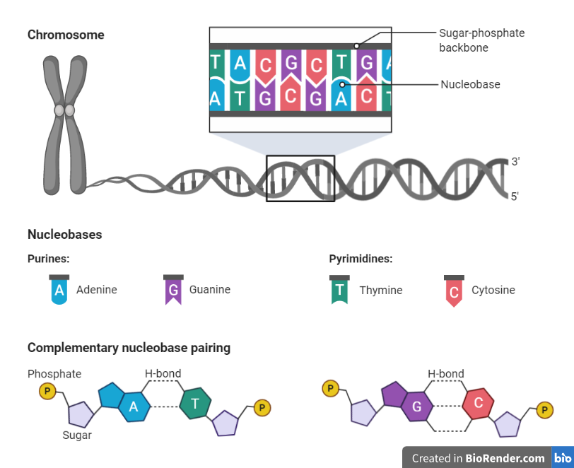
We are now aware of the components of the nucleotides as they were correctly predicted by Levene. These are a phosphate group, a pentose sugar molecule, deoxyribose in DNA and ribose in RNA, and a nitrogenous base. The bases that are found in DNA are adenine (A), thymine (T), guanine (G), and cytosine (C), while in RNA, there is uracil (U) instead of thymine with the other three remaining the same. These five bases are divided into two categories, purines, and pyrimidines. Adenine and guanine belong to purines, and they consist of two fused rings, while thymine, uracil, and cytosine consist of a single ring and are called pyrimidines.
Chargaff’s rules
A big step toward the discovery of the DNA structure was made by Erwin Chargaff, an Austrian biochemist. Chargaff studied the composition of DNA in different species to find whether it’s the same or not. A surprising finding was that in all the species he examined, the amount of adenine was similar to that of thymine, and the amount of guanine was similar to that of cytosine. This means that A=T and G=C. We can alternatively describe this as A+G=T+C, meaning that the number of purines is the same as the number of pyrimidines in the DNA of every species. However, the amount of each base, and consequently, that of its pair, varies among species. Therefore, the ratio A+T/G+C differs in the various organisms.
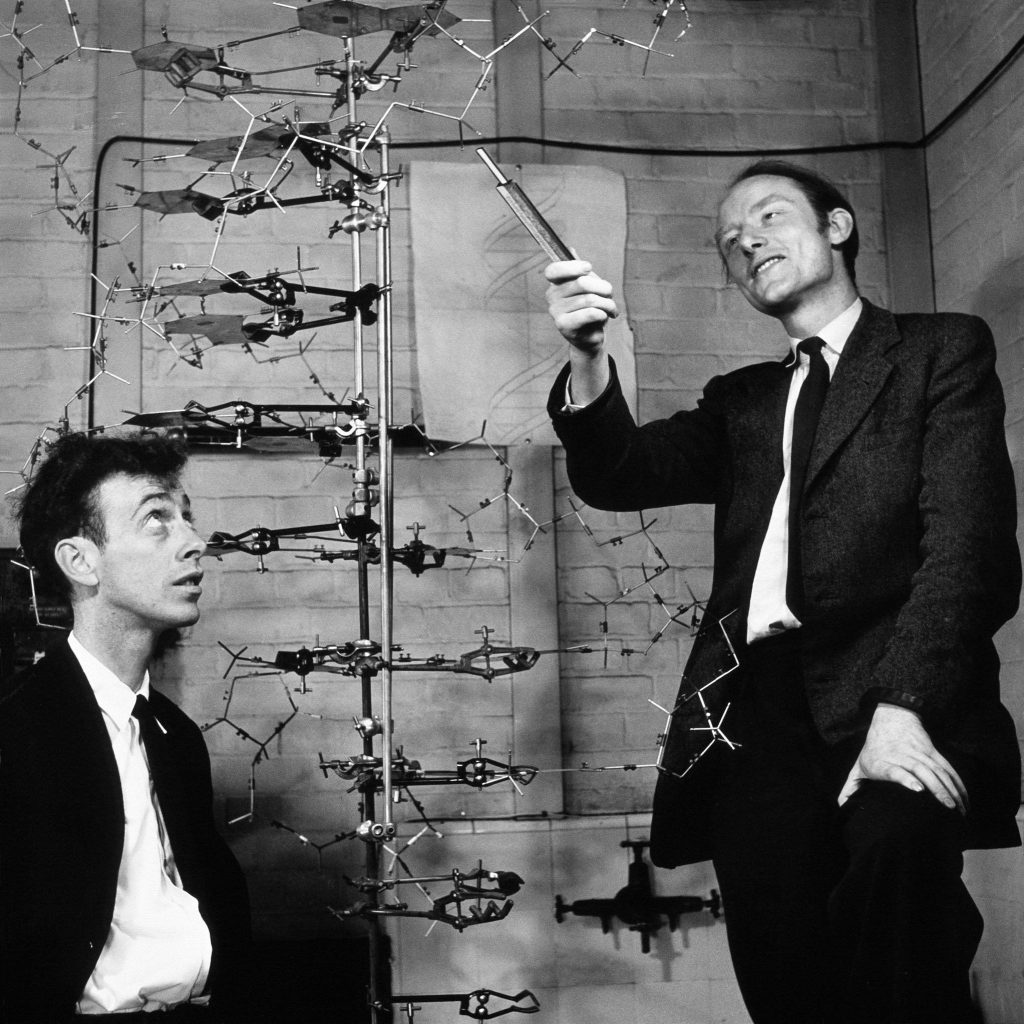
Watson, Crick, Wilkins, and Franklin
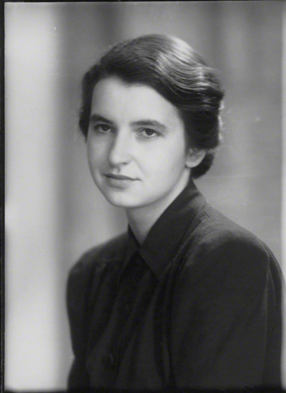
When all the above discoveries were combined, they contributed to the unraveling of the DNA structure. The final answer came from two teams. One of them was the team of American biologist James Watson and British physicist Francis Crick. The other one was that of physicist Maurice Wilkins and chemist Rosalind Franklin. Watson and Crick did not conduct any experiments of their own but used the results of previous studies to support their thoughts. They were especially based upon angle bonds and molecular distances that had been measured by Linus Pauling, an American biochemist that studied structural models.
Watson and Crick cut cardboards in the form of the DNA components and tried to arrange them in various ways to predict the three-dimensional structure of DNA. However, there were times when they could not find a correct way to put the pieces of the puzzle together due to a misunderstanding surrounding the atom configuration in some nitrogenous bases. When they shifted the positions of some of the atoms on these molecules, they managed to connect the bases in a way that confirmed Chargaff’s rule. Adenine and thymine were complementary bases that paired together, as was the case between guanine and cytosine. They also concluded that the base pairs were held together by hydrogen bonds.
Watson and Crick propose the Double Helix model
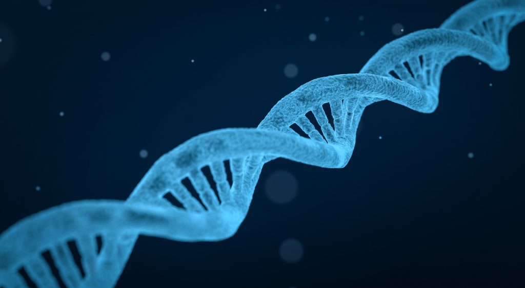
No matter their discoveries, they still had trouble arranging the whole picture of the molecule’s structure. Their questions, though, were eventually solved by the work of Franklin, the results of which were sent to Watson and Crick by Wilkins without her knowledge. Franklin was studying the structure of DNA by using a method called X-ray crystallography. This method uses X rays to create a picture of molecules. When X rays are directed to the crystal of a molecule, in this case of DNA, they hit its atoms. This interaction causes some rays to change direction and create a diffraction pattern, a picture, showing details of the arrangement of atoms in the molecule. Watching this image, Watson and Crick were able to understand how DNA is structured. They found that the nitrogenous bases are hidden on the inside of the molecule and interact with each other according to the complementary base-pairing rules, while the phosphate groups and the sugars create a sugar-phosphate backbone that is found on the outside. They also found that the DNA consisted of two strands that form a double helix that is also right-handed, due to the way the strands wrap around an imaginary axis.
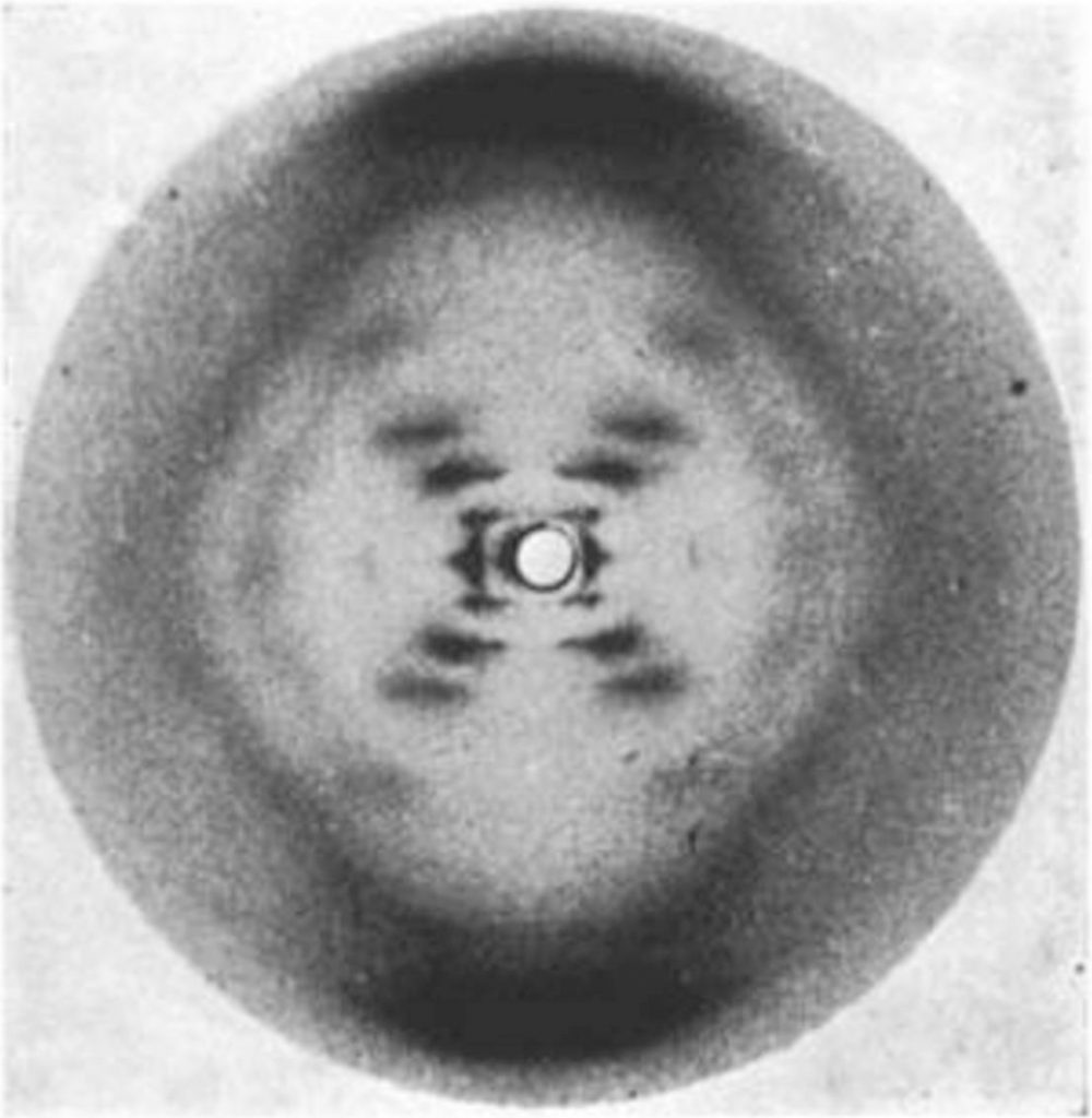
On the right: The X-ray diffraction photo of DNA from Rosalind Franklin (Image by Rosenfeld Media from Flickr under a Creative Commons Attribution 2.0 Generic License, no changes were made)
Watson and Crick discovered the 3D structure of DNA and published their findings in Nature on April 25th, 1953. The published results consisted of just one page and a drawing of the double helix drawn by Crick’s wife. Franklin died of ovarian cancer in 1958, and her contribution to the double helix discovery was not acknowledged. Watson and Crick were awarded the Nobel Prize in Medicine in 1962 and shared it with Wilkins without giving any credit to Franklin for her work.
Features of the double helix
Some of the structural features of the double helix as we know them today are given below:
- DNA consists of two polynucleotide strands that form a right-handed, double helix.
- On the outside of the molecule, there is a sugar-phosphate backbone, which is hydrophilic, while the nitrogenous bases are located on the inside and are hydrophobic. The bases follow the rules of complementary pairing (A pairs with T and G pairs with C). The two strands are held together by hydrogen bonds between the bases. There are two hydrogen bonds between A and T, and three hydrogen bonds between G and C.
- Every time, a purine (two-ringed structure) pairs with a pyrimidine (one single ring). Therefore, the distance between the bases is always the same, making the diameter of DNA around 2 nanometers.
- In each strand, the nucleotides bind one after the other with a covalent bond, called 3’-5’ phosphodiester bond. This bond joins the 3’ carbon atom of the sugar of the first nucleotide and the 5’ carbon atom of the following nucleotide.
- The two strands are antiparallel, meaning that they run alongside each other but in opposite directions. The 5’ end of one strand is located opposite the 3’ end of the other, and vice versa.
- The way the DNA molecule is structured drives its replication, and that was something Watson and Crick noticed shortly after they published their results. Due to the complementary base pairs, each DNA strand can act as a template for the synthesis of the other strand. This way, two new double helices are produced, and they are identical to each other and the original.
Sources:
https://www.nature.com/scitable/topicpage/discovery-of-dna-structure-and-function-watson-397/
https://www.history.com/this-day-in-history/watson-and-crick-discover-chemical-structure-of-dna