Chromosomes, chromatin, nucleosomes, genes, alleles? What is the difference between these terms? What are their roles? And how are they organized inside each one of the small cells of an organism? When you hear these words, you might think that they are pretty much the same, or you may not be able to distinguish their functions, but in this article, we will analyze and explain each of these terms and more.
The need for DNA packaging
We will not concern ourselves with prokaryotic organisms in this post, since they do not possess a nucleus and also the organization of their DNA is not very complicated. We are aware that eukaryotic organisms have cells that range in size and shape depending on the cell type or organism. However, in all cases, the common characteristic is that the nucleus is always very small compared to the size of the DNA it hosts inside.
If the nuclear DNA remained bare, uncondensed, with no modifications, it could reach as far as two meters in length, and it would absolutely not be able to fit inside the nucleus. Therefore, DNA needs to be condensed, with the assistance of proteins and enzymes to occupy less space. This state of DNA packaging is not permanent, though. During different phases of the cell cycle, there is a need for various levels of condensation because two basic functions must be ensured. One of them is, as we already mentioned, the packaging to fit in the cell. The other one is a need for certain regions to be accessible to cellular machines. What does this mean? For example, when a gene must be transcribed, transcription enzymes have to find the region of the gene and unwrap the double helix locally to synthesize an RNA molecule. That means that the gene must not be very tightly condensed. The same thing applies to replication. During replication, the entire DNA has to be gradually unwrapped to make copies of itself. Instead, when the cell divides, and there is no transcriptional activity, DNA must be tightly packaged to ensure a safe cell division.
Stages of chromatin condensation
Now let’s look at the process of DNA condensation from a bare molecule to a tightly-packed form. During interphase, DNA is less packaged. The cell is transcriptionally active, and that means that a large number of genes must be easily accessible to enzymes for transcription. DNA is always present in the form of a complex with proteins. This DNA-protein complex is called chromatin. Chromatin can have two different configurations, euchromatin, and heterochromatin. Euchromatin is the real (eu=real in Greek) chromatin, and this means it is loosely packed during interphase because it consists of active genes. Meanwhile, heterochromatin is densely packed because it consists of transcriptionally inactive regions.
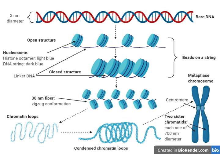
If DNA remained bare, its diameter would be only 2 nanometers, but it would also stretch to 2 meters length. Therefore, it follows packaging steps that make it grow in diameter but occupy less space in the nucleus. At first, a structure called nucleosome is formed when DNA wraps around a histone octamer. Histones are proteins evolutionarily conserved across species. Four of them take part in this step, H2A, H2B, H3, and H4. The histone octamer consists of eight molecules, two of each histone. Finally, 146 base pairs of DNA twist 1.65 times around a histone octamer creating a nucleosome. That happens multiple times, and a large number of nucleosomes are created one after the other, with a strand of linker DNA between consecutive nucleosomes. If you observe this chromatin stage in an electron microscope, you will see that it resembles beads on a string, which is an alternative name for this chromatin stage. The ‘beads on a string’ structure is furtherly condensed in a 30-nanometer fiber by coiling. Another histone called H1, along with the linker DNA takes part in this step.
However, the packaging process does not end there. Some proteins help the 30 nm fiber condense even more by creating loops. The presence of protein scaffolds makes these loops even tighter. Now the chromatin can reach a diameter of 700 nanometers. At this stage, the appearance of chromatin resembles the known structure of chromosomes that we see in books. So, chromosomes are the highest order of DNA organization. Don’t forget that by now, DNA has been replicated. That means there is double the information inside the nucleus. Each chromosome consists of two identical DNA molecules that have occurred from replication. These are called sister chromatids and remain linked to one another with the help of a structure called the centromere. Chromosomes appear at their most compacted form during metaphase, a specific stage of mitosis, when they consist of two sister chromatids, each one with a width of 700 nm. Therefore, their total diameter is about 1400 nanometers.
Number of chromosomes and karyotype
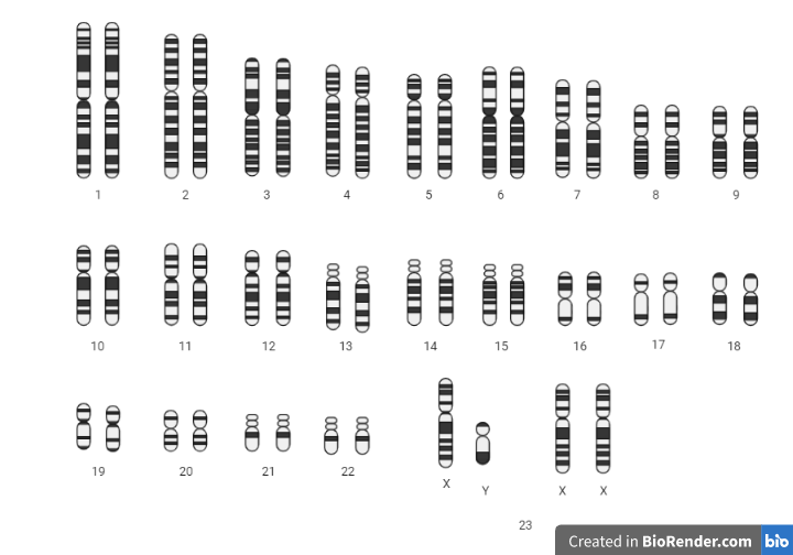
In a previous post, we talked about the differences between somatic cells and gametes. The first are diploid cells, and the latter are haploid cells. However, the number of chromosomes differs among species. For example, humans have 46 chromosomes in their somatic cells, while horses have 64, and mice have 40. We will focus on the case of humans, but the same applies to other organisms as well. Of the 46 chromosomes, 44 are called autosomes, and the other 2 are the sex chromosomes. Autosomes carry information that determines characteristics of the person’s somatic cells, while sex chromosomes determine the sex, although they have some other functions as well. Females possess two X chromosomes, while males possess one X and one Y chromosome. Half of the genetic information of a somatic cell derives from the person’s mother and the other half from their father. Therefore, of the 46 chromosomes, there are 23 maternal chromosomes and 23 paternal ones. Of the 23 chromosomes, 22 are autosomes, and one is a sex chromosome. A mother always contributes an X chromosome to her offspring, while a father can pass down either an X or a Y chromosome. Since gametes are haploid cells, they contain half the genetic information of the somatic cells, meaning 23 chromosomes (22 autosomes and one sex chromosome).
Chromosomes can be observed under a microscope after they’ve been isolated from a person’s cells. Metaphase chromosomes are best visible due to their high levels of condensation, as we saw earlier. Dyes that stain the chromosomes are used to make it easier to classify them in pairs. The image of chromosomes arranged in pairs, from largest to smallest, is called a karyotype. By looking at the karyotype, we can recognize any abnormalities considering the number and size of the chromosomes. In the case of humans, a normal karyotype consists of 23 pairs of homologous chromosomes. Homologous chromosomes are two pieces of DNA that carry the same genes in the same regions. One chromosome of each pair originates from the father and the other one from the mother. The two X chromosomes of a female are homologous, whereas X and Y chromosomes in males contain only a few homologous genes, and therefore, are considered non-homologous.
Homologous chromosomes, genes, and alleles
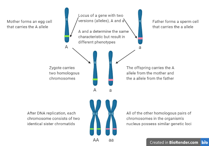
Genes are the functional unit of chromosomes. They are nucleotide segments that code for proteins, which determine our characteristics. We know that genes are scattered along the whole genome of an organism, but not all individuals possess the same versions of each gene. Homologous chromosomes have specific genes in the same region, called genetic locus. Variations in these genes are called alleles, and they code for slightly altered versions of the gene product. A characteristic might be determined by multiple genes or by just one. And each of these genes might occur with two or more alleles. Let’s talk about the simple case of a characteristic that is controlled by one gene that has two alleles, A and a, which code for two different proteins. Each person has two of these alleles that can be AA, Aa, or aa. One of them is located on one chromosome, and the other one is located in the same locus on the homologous chromosome. The two alleles can be the same (AA or aa) or different (Aa) depending on the alleles the person has inherited from their two parents.
For more information:
https://www.ncbi.nlm.nih.gov/books/NBK21500/
Sources:
https://www.nature.com/scitable/topicpage/chromosomes-14121320/ https://openoregon.pressbooks.pub/mhccmajorsbio/chapter/dna-organization-inside-a-cell/